Advanced Dental Technology: Precision, Safety, and Comfort
Integrating High-Tech Innovation with Holistic Care
At Smile Angels of Beverly Hills, we believe that world-class dental care requires more than just a gentle touch; it demands the highest level of diagnostic precision. Dr. Bruce Vafa has invested in state-of-the-art dental technology to ensure your treatments are minimally invasive, safer, and more effective than traditional methods.
For our holistic patients, this technology is critical. It allows us to:
Minimize Radiation: Using advanced digital imaging.
Eliminate Guesswork: Detecting decay before it becomes visible to the naked eye.
Improve Biocompatibility: Using your body’s own materials for healing.
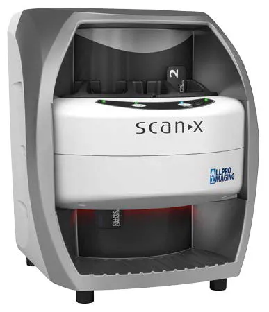
ScanX Duo: Ultra-Low Radiation Digital X-Rays
Your long-term health is our priority. That is why we have replaced old-fashioned film X-rays with the ScanX Duo Digital Imaging System.
90% Less Radiation: Drastically reduces your exposure compared to traditional film.
Instant Clarity: Digital images appear instantly on our screens, allowing Dr. Vafa to zoom in and diagnose issues with pinpoint accuracy.
Comfortable Plates: We use flexible phosphor plates that are thin and wireless, eliminating the discomfort of biting down on rigid film holders.
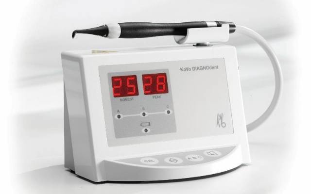
DIAGNOdent: Laser Cavity Detection
Traditional metal probes (poking and prodding) are often uncomfortable and can miss up to 50% of hidden decay. Dr. Vafa utilizes DIAGNOdent, a revolutionary laser fluorescence system that scans your teeth for hidden cavities.
How it Works: The laser measures the reflection of light within the tooth structure. Healthy teeth reflect light differently than teeth with decay.
The Benefit: We can detect “micro-cavities” years before they appear on X-rays. This allows for micro-fillings that preserve almost all of your natural tooth structure, preventing the need for large drills or crowns later.
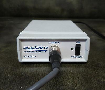
Acclaim® Intraoral Camera
We believe in total transparency—you see what we see. The Acclaim Intraoral Camera allows Dr. Vafa to show you high-definition, real-time images of your teeth on a monitor. This ensures you fully understand your oral health needs and can make informed decisions about your treatment.
Epic 10 Diode Laser: Soft Tissue & Pain Therapy
Lasers have transformed holistic dentistry by reducing the need for sutures and anesthesia. Dr. Vafa uses the Epic 10 Diode Laser for a variety of comfortable treatments:
Gum Therapy: Vaporizes harmful bacteria below the gumline to treat periodontal disease without cutting.
Pain Management: The laser’s therapeutic wavelength can be used to reduce inflammation and pain associated with TMJ disorders and facial tension.
Teeth Whitening: Provides faster, more effective whitening with less sensitivity than traditional gels.
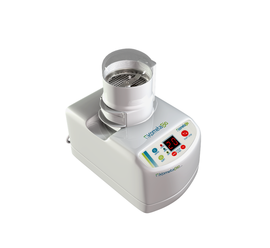
Smart Dentin Grinder: The Gold Standard in Bone Grafting
For patients requiring dental implants, bone quality is everything. In the past, bone grafts required using synthetic materials or bone from cadavers/animals. Dr. Vafa is one of the few dentists to offer the Smart Dentin Grinder.
100% Autologous (Your Own Body): If you have an extraction, we can process that extracted tooth into high-quality bone graft material in just minutes.
Faster Healing: Because the graft is made from your own dentin (which is chemically similar to bone), your body accepts it immediately, often cutting healing time in half (3 months vs. 6+ months).
Holistic Purity: No risk of disease transmission or rejection, as the material comes entirely from you.
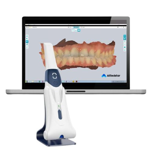
Medit i500 Intraoral Scanner
Say goodbye to the gag-inducing, messy putty used for dental impressions. Dr. Vafa uses the Medit i500, a specialized 3D scanner that captures a digital replica of your teeth in seconds.
Superior Accuracy: Digital scans are micron-accurate, ensuring your crowns, veneers, and Invisalign aligners fit perfectly the first time.
Patient Comfort: The slim scanning wand is non-invasive and fast, making it ideal for patients with a strong gag reflex.
Experience the difference of a dentist who listens, cares, and excels.
Whether you are looking for a holistic approach, relief from jaw pain, or a cosmetic smile makeover, you are in expert hands.
Schedule YOUR CONSULTATION
Come in for a consultation and find out all about the best dentistry experience and cutting edge technology in Beverly Hills.
By submitting this, you agree to be contacted by us. For more information, [lease read our privacy policy